IPA 2023 Photogenic Science Contest Winners
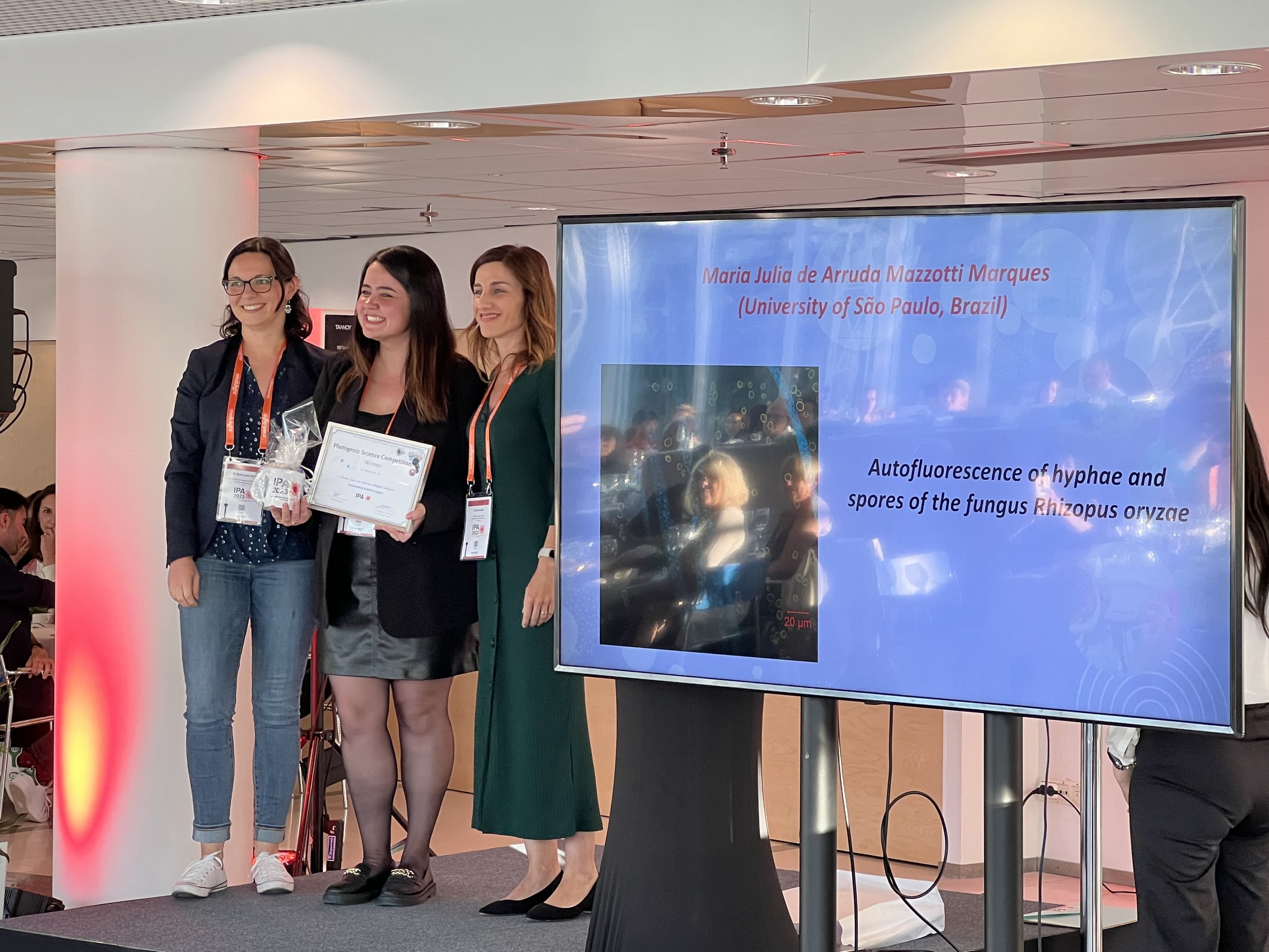
Category: Fundamental Science
Maria Julia de Arruda Mazzotti Marques, University of Sao Paulo, Brazil
Maria Julia de Arruda Mazzotti Marques
University of Sao Paulo, Brazil
Title: Autofluorescence of hyphae and spores of the fungus Rhizopus oryzae
Winner: Maria Julia de Arruda Mazzotti Marques
PhD student at University of Sao Paulo, Brazil
Image description: During the COVID-19 pandemic, several complications arose in infected patients, and one of them was mucormycosis, an extremely aggressive fungal disease with a high mortality rate, especially in people with compromised immune systems. The majority of mucormycosis cases are caused by the fungus Rhizopus oryzae, also known as black fungus.
© 2023 Maria Julia de Arruda Mazzotti Marques
Autofluorescence of hyphae and spores of the fungus Rhizopus oryzae
In October 2022, the WHO released the first list of fungi that pose a major public health risk due to the lack of rapid diagnostics and the invasive way these fungal diseases affect debilitated patients. One of the fungi mentioned in this list is Rhizopus spp., which causes mucormycosis. The WHO aimed to publicize this list to draw the world's attention to the importance of investing in research and developing new methods of diagnosis and control for these infections [1].
The treatments used are based on high doses of amphotericin B and posaconazole, along with surgical resections when possible. However, even with aggressive antifungal treatment, the estimated attributable mortality rate is high. In the absence of surgical debridement of infected tissue, antifungal treatment alone is not curative. Therefore, there is a need for the development of adjunctive treatments. Antimicrobial photodynamic therapy (aPDT) may be an auxiliary therapeutic option for mucormycosis.
Due to the lack of reports in the literature on the morphology and photodynamic inactivation of R. oryzae, we performed a characterization of this fungus using Confocal Fluorescence Microscopy. The image shows the autofluorescence of the fungus R. oryzae, through which it is possible to observe the natural fluorescence of hyphae and spores. It is noted that the hyphae are cylindrical and quite elongated, and regions with no fluorescence in the hyphae are observed; these regions likely contain melanin and are highly light absorbing.
References:
[1] WORLD HEALTH ORGANIZATION. WHO fungal priority pathogens list to guide research, development and public health action. Disponível em: https://www.who.int/publications/i/item/9789240060241
Category: Pre-Clinical Science
Maria Rosada, University College London, UK
Maria Rosado
University College London, UK
Title: Shine bright like a diamond
Winner: Maria Rosado
Research Assistant, University College London, UK
Image description: Here is an image captured through confocal microscopy of U87 cells, a type of human glioblastoma. The image depicts the co-localization of two proteins - WIP (in green) and IQGAP (in blue) - along with actin filaments (in red). Thus, we can ensure the location of the two proteins.
The actin cytoskeleton stands as a crucial and intricate structural framework within cells, orchestrating a range of fundamental processes that encompass cell mobility and configuration. This intricate framework is composed of a complex interconnection of actin polymers, intricately interwoven to create a dynamic network. Notably, the intricate composition of the actin cytoskeleton involves the active participation of various proteins, with prominent examples including the likes of WASP (Wiskott Aldrich Syndrome Protein) and WIP (WASP-interacting protein). These proteins exert pivotal functions in the orchestration and reinforcement of this cytoskeletal matrix, ensuring its structural organization and integrity.
© 2023 Maria Rosado
Shine bright like a diamond
Of particular emphasis in the presented image is the WIP protein, visually indicated in green. This specific protein serves as a focal point due to its remarkable role in modulating the stability and operational dynamics of the WASP protein, denoting a vital aspect of cytoskeletal dynamics (1). It’s important to note that the interaction between WIP and WASP plays a significant role in shaping cellular responses and behaviors. Adding to its significance, WIP expression demonstrates an intricate link to various types of cancer. In the context of lymphomas, WIP functions as a tumor suppressor, acting to restrain the progression of malignancies. Conversely, in the realm of solid tumors, WIP takes on a different role, behaving as an oncogene and potentially contributing to the initiation and advancement of tumorigenic processes.
The primary aim of this study was to determine the natural placement of WIP within human glioblastoma cells. Additionally, it aimed to verify its co-localization with the IQGAP protein (in blue), previously identified in its interaction network.
Gliomas are abnormal growths within the central nervous system that impact both the brain and spinal cord. Within this category, there are diffuse gliomas, which lack distinct boundaries and are recognized for being the prevalent form of primary malignant growths. Glioblastoma is a specific subtype belonging to the group of diffuse gliomas (2). There are different grades of glioblastomas, with grade four being the most aggressive, also known as glioblastoma multiforme (GBM) (3).
Using confocal microscopy to capture such images, we can precisely determine the positioning of WIP within both the cell nucleus and the cytoplasm. This is particularly evident in the outer regions of the cell, adjacent to the actin filaments (in red), where IQGAP is also present. As a result, we successfully verified the shared presence of these two proteins in glioblastoma cells. Our primary objective remains centered on furnishing novel insights into the mechanisms underlying tumors associated with these proteins. This information is invaluable in devising innovative therapeutic approaches.
References:
1. Antón, I. A., Jones, G. E., Wandosell, F., Geha, R. y Ramesh, N. (2007). WASP interacting protein (WIP): working in polymerisation and much more. Trends in Cell Biology, 17(11), 555-562.
2. Ferris, S. P., Hofmann, J. W., Solomon, D. A. y Perry, A. (2017). Characterization of gliomas: from morphology to molecules. Virchows Archiv, 471, 257-269.
3. Holland, E. C. (2000), Glioblastoma multiforme: The terminator. Proceedings of the National Academy of Sciences of the United States of America, 97(12), 6242-6244.
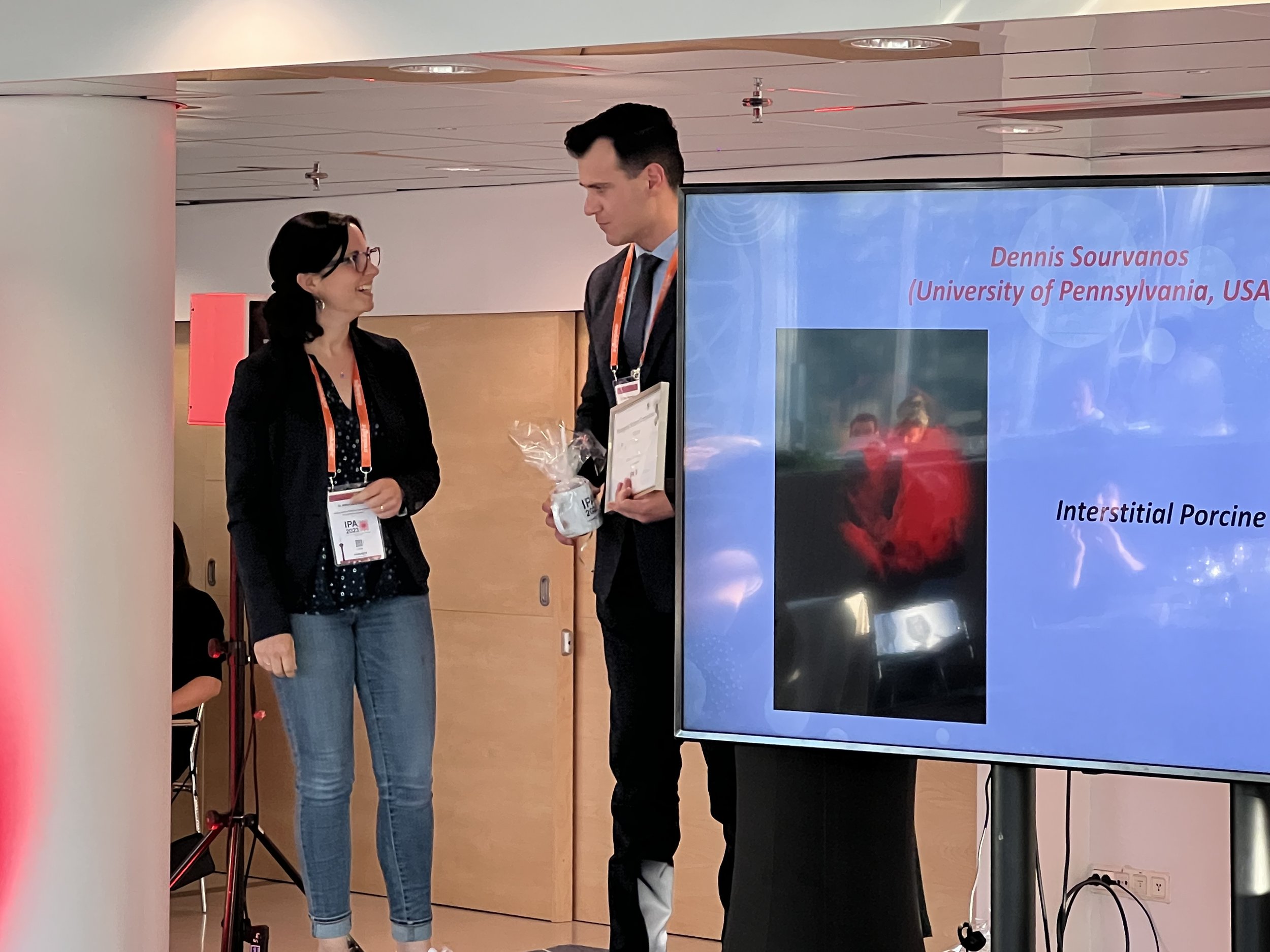
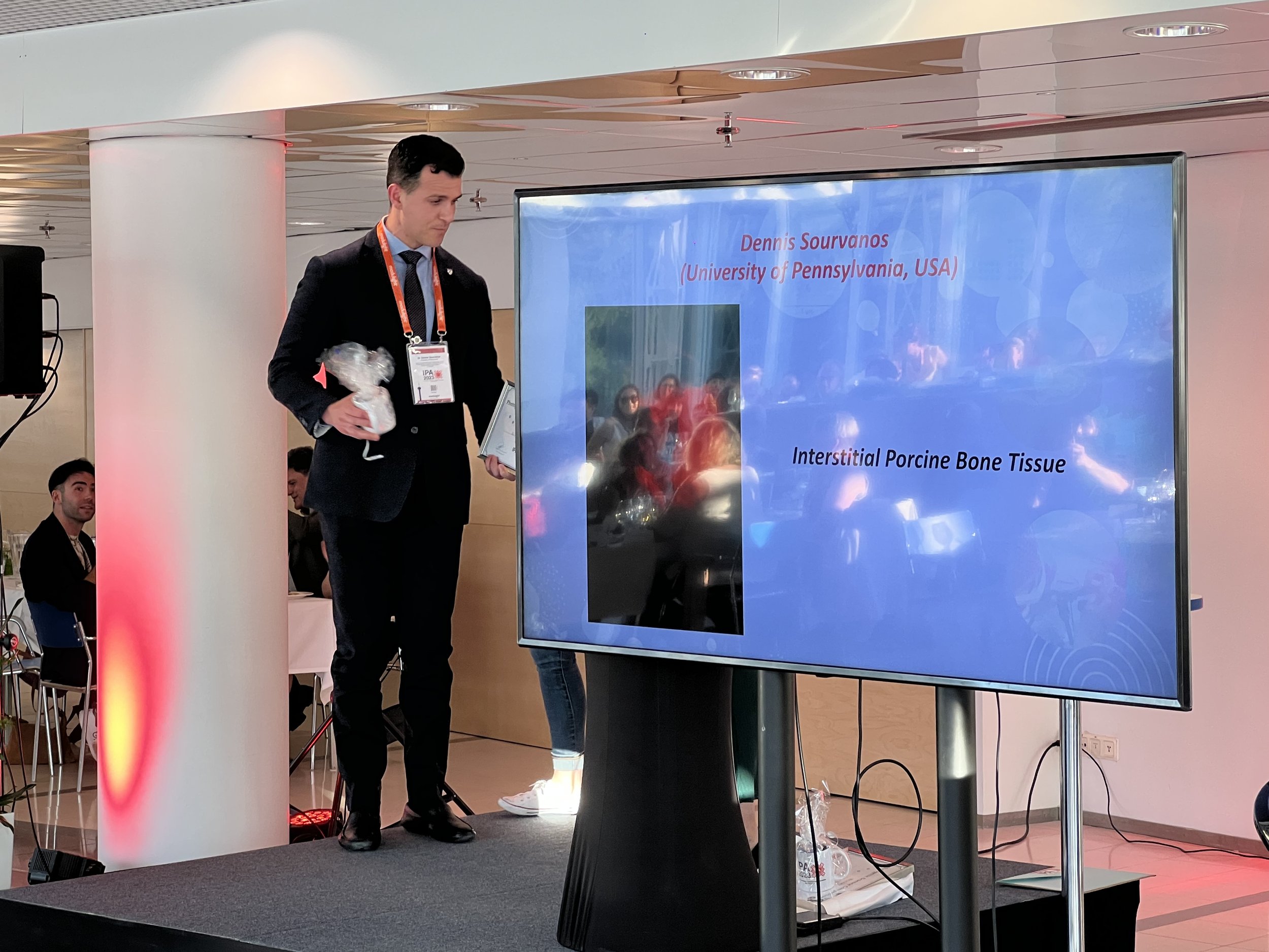
© 2023 Dennis Sourvanos
Interstitial Porcine Bone Tissue
Category: Clinical Science
Dennis Sourvanos
University of Pennsylvania, USA
Title: Interstitial Porcine Bone Tissue
Winner: Dennis Sourvanos
Post-doctoral fellow, University of Pennsylvania, USA
Image description: The 660nm Red Wavelength point light source is illuminated through porcine bone tissues elegantly unraveling absorption and scattering coefficients. This image unveils a resplendent point light source gracefully piercing the cortical bone tissue of the porcine condyle. Two 4cm parallel osteotomies accommodate a light source and an isotropic detector, which is connected to a motorized dosimetry system.
© 2023 Marvin Xavierselvan
Spheroid Viability Assessment: Live/Dead Imaging Reveals the Impact of Photodynamic Therapy
Category: People’s Choice
Marvin Xavierselvan
Tufts University, USA
Title: Spheroid Viability Assessment: Live/Dead Imaging Reveals the Impact of Photodynamic Therapy
Winner: Marvin Xavierselvan
PhD student at Tufts University, USA
Image description: The image showcases the assessment of spheroid viability following photodynamic therapy (PDT). DAPI staining visualizes the nuclei of all cells, while live cells are highlighted using Calcein-AM staining. Dead cells are specifically labeled with EthD-1 staining. The staining comprehensively evaluates treatment efficacy and cell viability in response to PDT.
The oxygen dependency of photodynamic therapy (PDT) presents a significant challenge in cancer treatment due to the prevalent hypoxia within the cores of solid tumors. The low oxygen content imparts tumors with resistance to other major therapies. To better understand the relevance of hypoxia in cancer treatment, it is vital to develop a suitable model that mirrors tumor oxygen and nutrient gradient, cellular heterogeneity, tumor architectures, and physiological conditions. While 2D in vitro models enable controlled experiments to test potential therapeutic strategies, they do not provide the same level of cellular heterogeneity and tumor architectures as in vivo settings. Conversely, in vivo experimentation is resource-intensive and lacks high screening throughput. Multicellular tumor spheroids models bridge this gap and emulate the tumor microenvironment and hypoxic conditions.
The 3D spheroids models hold immense promise, as they offer a closer representation of the physiological conditions found within real tumors. Their unique advantage lies in capturing intricate cell-cell and cell-extracellular matrix interactions, mirroring the complex dynamics of actual tumor microenvironments. By harnessing this model, we can study the impact of hypoxia on various cellular processes and test potential therapeutic strategies in controlled and relevant settings.
We employed this model to create hypoxic head and neck cancer (HNC) spheroids and monitored the development and progression of hypoxia. Following the hypoxia characterization, we assessed the photosensitizer (BPD) uptake and spatial distribution in HNC spheroids. Finally, we treated the spheroids with BPD-PDT and assessed the viability using live-dead staining. Calcein-AM stains the live cells while EthD-1 stains dead cells specifically. DAPI staining was performed to visualize the nuclei of all cells. The staining comprehensively evaluates the treatment efficacy and cell viability of spheroids in response to PDT. Overall, the spheroids model can shed light on the interplay between hypoxia and PDT efficacy and can contribute to the development of more effective cancer therapies.
Photogenic Scientific awards presented at the 18th IPA World Congress in Tampere, Finland.


